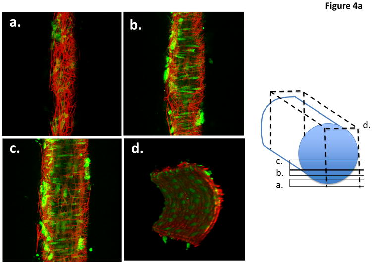
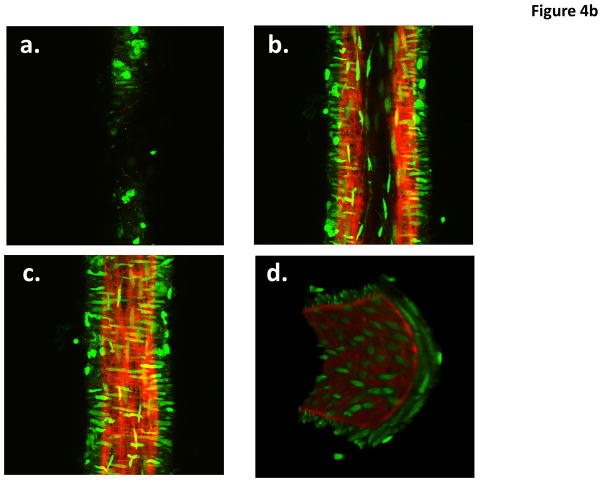
Figure 4.
Example pseudocolored images of a cannulated and pressurized cremaster arteriole (A) and a similarly prepared small cerebral artery (B). The images represent adventitial (Panel a), mid-wall (Panel b) intimal (Panel c) and end view (Panel D) sections. Vessel segments were fixed while pressurized (see text for details) and stained with Alexa 633 (for ECM structures) and Yo-Pro-1 iodide (nuclei). Images are representative of n = 11 (cremaster) and 6 (cerebral). Movie files for the complete image stacks and 3D rotating representations are shown in the Supplementary Material.


