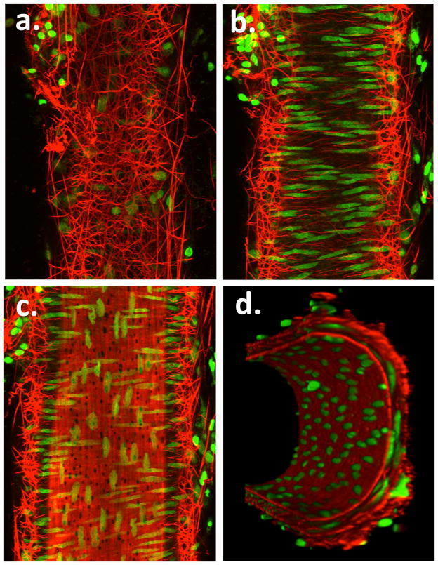
Figure 6.
Example pseudocolored images showing adventitial (Panel a), mid-wall (Panel b) intimal (Panel c) and end view (Panel d) sections of a small cannulated mesenteric artery. Vessel segments were fixed while pressurized and stained with Alexa 633 (for ECM structures) and Yo-Pro-1 iodide (nuclei). Images are representative of n = 3 vessels. Movie files for the complete image stacks and 3D rotating representations are shown in the Supplementary Material.

