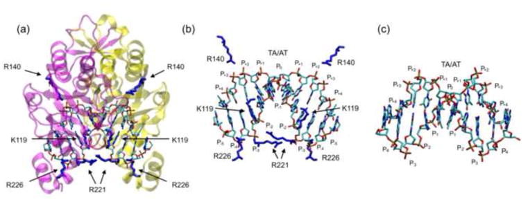
Figure 1. The EcoRV-DNA-Ca2+ complex.
(a) Location of the eight basic residues (4 per monomer) that contact the phosphate backbone in the specific complex (PDB ID 1B94)60. The protein backbone is in magenta and yellow (to distinguish the subunits) and cationic side chains that contact DNA phosphates are in dark blue. (b) Magnification of the DNA from the complex with protein backbone removed for clarity. Basic side chains that contact the phosphate backbone are shown in blue. The DNA bends downward and phosphate positions on each DNA strand are numbered as in Fig. 2a. (c) Conformation of free CCGATATCGG (PDB ID 1ZFC)105 for comparison. Molecular images in all figures were created with the molecular graphics program “VMD”106.

