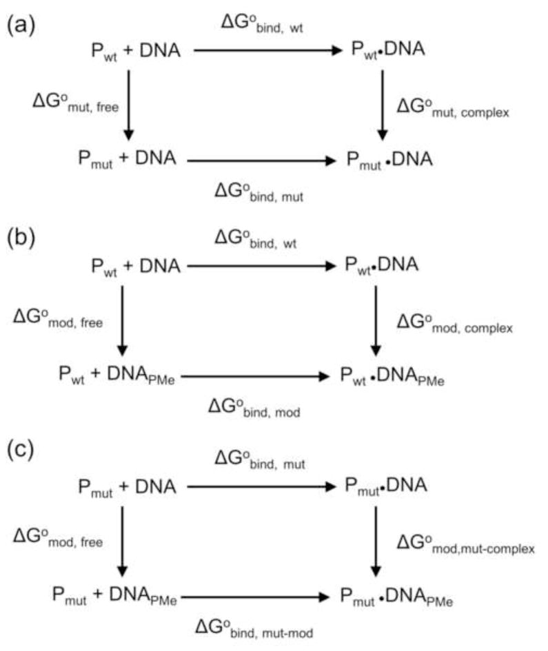
Figure 3. Thermodynamic pseudocycles.
(a) Equilibrium binding of unmodified DNA to wild type and mutant EcoRV; (b) Equilibrium binding of wild type EcoRV to unmodified and PMe modified DNA; (c) Equilibrium binding of mutant EcoRV to unmodified and PMe modified DNA. The free energy changes (ΔG°) for the reversible processes indicated by horizontal arrows can be determined by equilibrium binding experiments, but the “processes” indicated by vertical arrows represent chemical modifications or protein mutations, with free energy changes that are not experimentally accessible.

