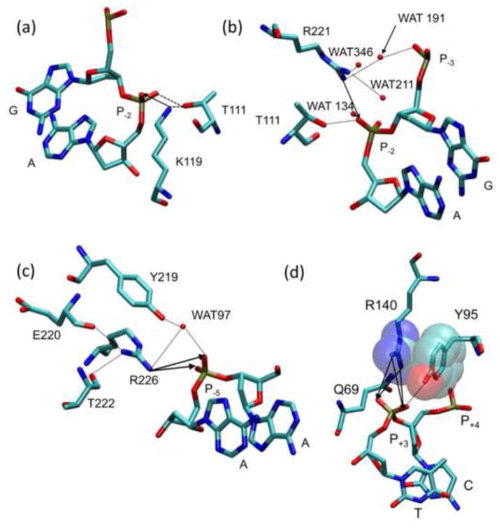
Figure 4. Interactions of cationic sidechains with DNA phosphates.
Contacts made to the (a) K119, (b) R221, (c) R226, and (d) R140 side chains in crystal structure PDB ID 1B94. Neighboring side chains, bases, and water molecules are labeled and atoms are represented by color (C = cyan, O = red, N = blue, and P = brown). Hydrogen bonds and Coulombic interactions are represented by dotted lines and arrows respectively. (d) Van der Waals interactions between the R140 guanidino and the Y95 phenyl are shown in a semi-transparent space fill representation.

