
Figure 9.
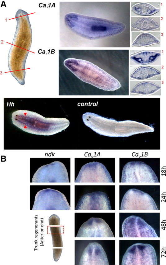
Localization of Cav1 channels with Hh in the planarian nervous system. A, Top, Cav1A and Cav1B localization in whole-mount and sectioned samples. Brightfield image of intact planarian shows location of cross sections: anterior (1, top), pharyngeal (2, middle), and postpharyngeal (3, bottom). Sections are orientated with the ventral side at the bottom. Bottom, Whole-mount in situ hybridization of Hh showing localization of mRNA within the ventral nerve cords (red arrows) compared with controls. B, Whole-mount in situ hybridization of ndk (a brain marker), Cav1A, and Cav1B in the anterior blastema during trunk fragment regeneration at the indicated times.