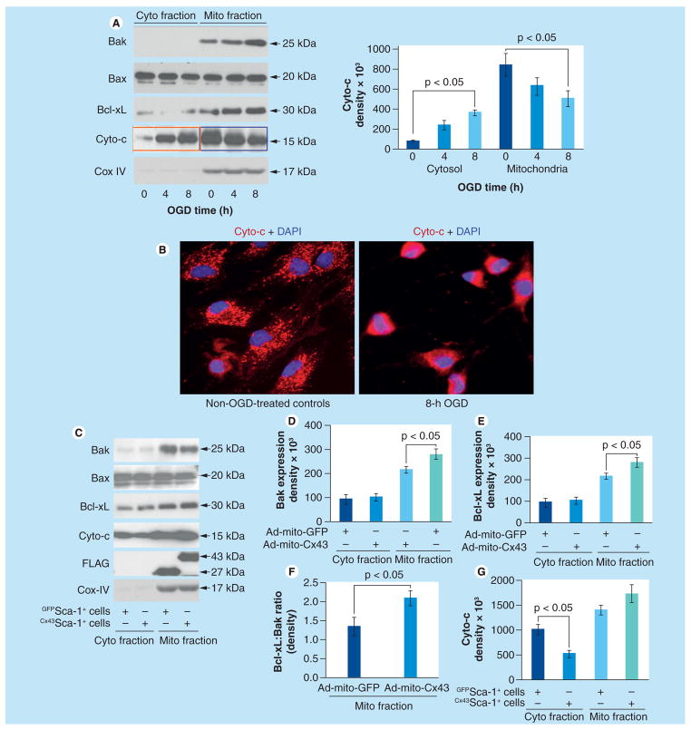
Figure 3. Mitochondrial connexin-43 alters dynamics of antiapoptotic Bcl-2 family members.
(A) Western blotting using cytoplasmic and mitochondrial protein fractions from Cx43Sca-1+ cells after OGD treatment for 0, 4 and 8 h. Increasingly higher expression of Bak, Cyto-c and Bcl-xL was observed in mitochondrial fractions at 4 and 8 h of OGD treatment as compared with cytoplasmic fractions. Cox IV was used as a loading control and to show the purity of cytoplasmic and mitochondrial fractions. (B) Fluorescence immunostaining of Sca-1+ cells exposed to 8 h OGD showed loss of punctuate distribution of Cyto-c. The nuclei were visualized with DAPI staining (blue; original magnification ×40). (C–G) Western blots of cytoplasmic and mitochondrial protein fractions from Cx43Sca-1+ cells and GFPSca-1+ cells after 8 h OGD treatment showed that Bax expression in both mitochondrial and cytoplasmic fractions remained unchanged. However, Bcl-xL expression increased significantly in mitochondrial fractions of Cx43Sca-1+ as compared with GFPSca-1+ cells, whereas expression of both Bak and Bcl-xL in cytoplasmic fractions of Cx43Sca-1+ cells remained unchanged. The Bcl-xL:Bak ratio was significantly higher in mitochondrial fractions of Cx43Sca-1+ cells. These molecular events significantly favored reduced Cyto-c release into cytosolic fractions of Cx43Sca-1+ cells. Cox IV was used as a loading control and to show the purity of cytoplasmic and mitochondrial fractions. The graphs show band intensity from three independent experiments.
Ad: Adenoviral; Cx43: Connexin-43; Cyto: Cytoplasmic; Cyto-c: Cytochrome-c; Mito: Mitochondrial; OGD: Oxygen–glucose deprivation.

