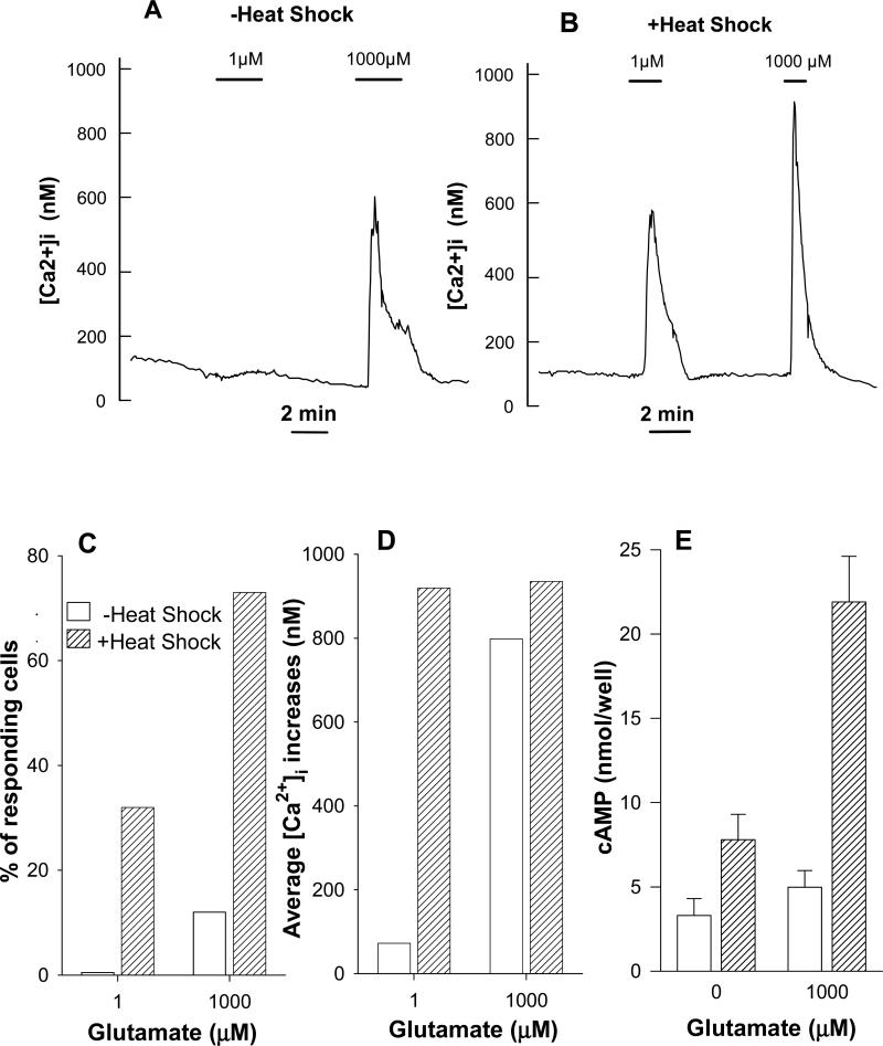
Fig. 6.
In mGluR1a transfected CHO cells heat shock increases both agonist potency and proportion of cells responding to agonist. (A) Average Ca2+ signals of responding cells stimulated by glutamate under control conditions (10 responding cells of 82 in the field of observation). (B) Cells were heated at 42.5°C for 2 hrs 1 day before measurements were performed (27 responding cells of 51 in the field of observation). Data of two representative experiments are shown; similar results were obtained in 3-5 other experiments. (C) Summarized data indicating proportion of cells responding to different glutamate concentrations under control conditions (17 out of 222 cells in 3 independent experiments) and after heating (141 out of 243 cells in 5 independent experiments). (D) Average [Ca2+]i signals of the same (C) responding cells. (E) Heat shock enhances cAMP formation in mGluR1a expressing CHO-K1 cells. Cells were heated for 2 hours at 42.5°C. After additional 24 hours cells were used for determination of cAMP accumulation stimulated by agonist in presence of phosphodiesterase inhibitor IBMX.

