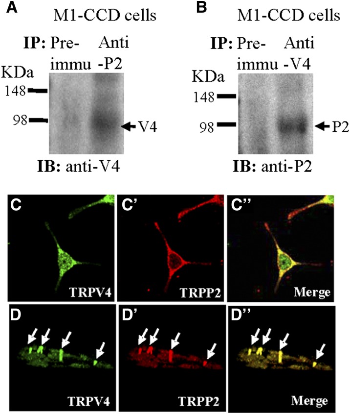
Figure 3.
Physical interaction between TRPV4 and TRPP2 in M1-CCD cells. (A and B) Coimmunoprecipitation. The pulling antibody and the blotting antibody are indicated. Control immunoprecipitation was performed using preimmune IgG (labeled as Preimmu). Anti-P2 indicates anti-TRPP2, anti-V4 indicates anti-TRPV4, IB indicates immunoblot, and IP indicates immunoprecipitation (n=3 experiments). (C–D″) Double immunostaining. (C and D) TRPV4 and (C′ and D′) TRPP2 were colocalized in (C″ and D″; merge) the cytosol, the plasma membrane, and the primary cilium of M1-CCD cells. D–D″ are confocal images of z-series stacks showing the primary cilium emerging from the apical membrane.

