Figure 4.
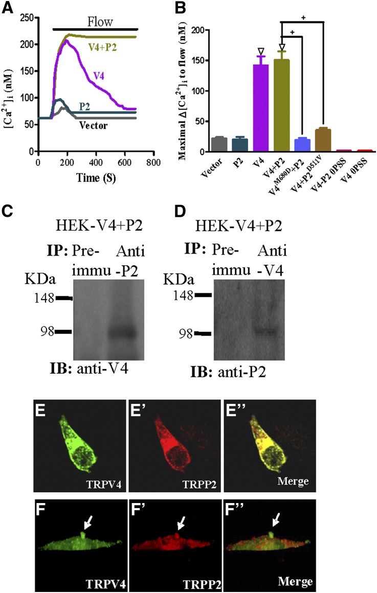
Physical association and functional role of TRPV4 and TRPP2 in HEK293 cells that were coexpressed with TRPV4 and TRPP2. (A and B) Flow-induced [Ca2+]i responses. (A) Representative traces illustrating the time course of flow-induced [Ca2+]i responses. (B) Summary showing the maximal change in flow-induced [Ca2+]i responses (Δ[Ca2+]i). Cells were bathed in NPSS containing 1% BSA except for 0PSS series (labeled bars in B), in which the bath was Ca2+-free. Cells were transfected with an empty vector (vector), TRPP2 (P2), TRPV4 (V4), TRPV4+TRPP2 (V4+P2), TRPV4M680D+TRPP2 (V4M680D+P2), or TRPV4+TRPP2D511V (V4+P2D511V). Data are given as the mean ± SE (n=6–8 experiments and 8–20 cells per experiment). ΔP<0.01 versus vector. +P<0.01 versus V4+P2. (C and D) Coimmunoprecipitation. The pulling antibody and the blotting antibody are indicated. Control immunoprecipitation was performed using the preimmune IgG (Preimmu; n=3 experiments). (E–F″) Double immunostaining. (E and F) TRPV4 and (E′ and F′) TRPP2 were colocalized in (E″ and F″; merge) the cytosol, the plasma membrane, and the primary cilium of transfected HEK293 cells. F–F″ are confocal images of z-series stacks showing the primary cilium emerging from the apical membrane.

