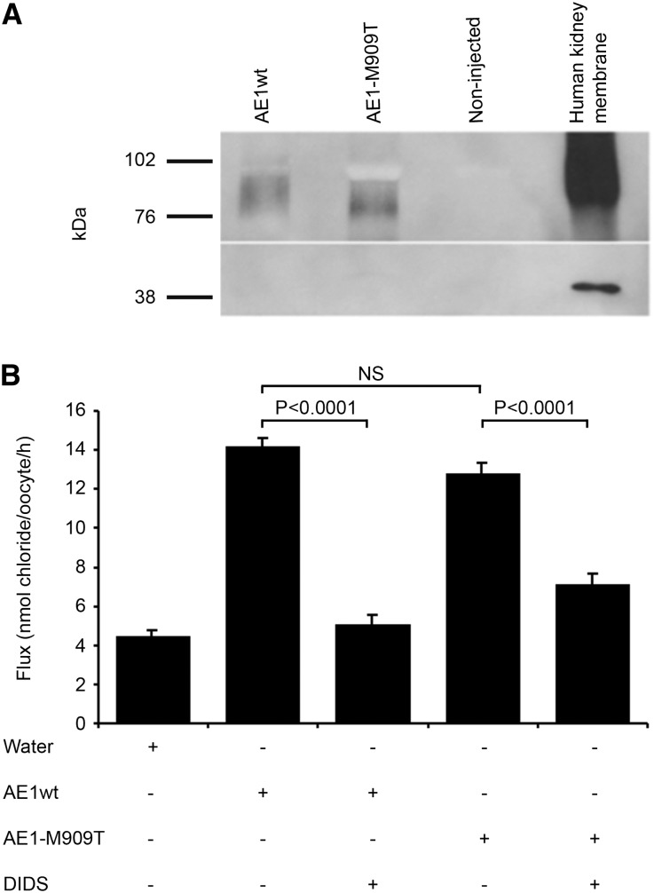
Figure 2.
Expression of kAE1 in Xenopus oocytes. (A) Western blot analysis of microinjected Xenopus oocytes confirms similar levels of surface expression of kAE1wt and kAE1-M909T (approximately 95 kDa biotinylated surface fraction) using the anti-AE1 monoclonal antibody Bric-170. Actin control confirms biotinylation of surface fraction only. (B) 36Cl uptake by oocytes injected with wild-type (WT) or mutant (M909T) kAE1 cRNA in the presence or absence of DIDS. Control oocytes were injected with water. Values are means ± SEM of influx pooled from five separate experiments with 9–33 oocytes/group. NS = not significant.

