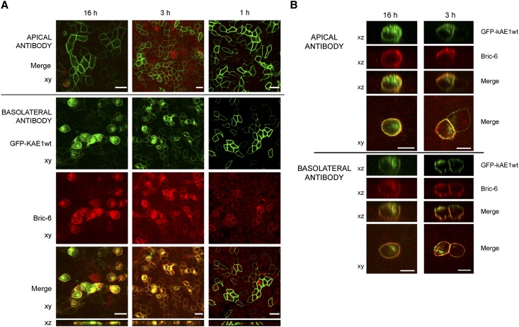
Figure 5.
Antibody uptake assay showing that kAE1wt traffics directly to the basolateral membrane. After induction of GFP-kAE1 expression, Bric-6 antibody was added to either the apical or basolateral compartment immediately (16 h) or at a later time (3 or 1 h) before fixing. (A) In cells expressing GFP-kAE1wt, no staining was evident with apical application of antibody (top panel), but basolateral application led to labeling of GFP-kAE1wt and internalization of the antibody–AE1 complex, with intracellular accumulation most evident in the 16-h assay (left column). (B) In cells expressing GFP-kAE1-M909T, application of Bric-6 to either compartment labeled both apical and basolateral surface kAE1-M909T, with increased staining at the opposing surface evident with longer incubation times (16 versus 3 h). Representative of (A) seven or (B) two separate experiments. Scale bars, 20 µm in A; 10 µm in B.

