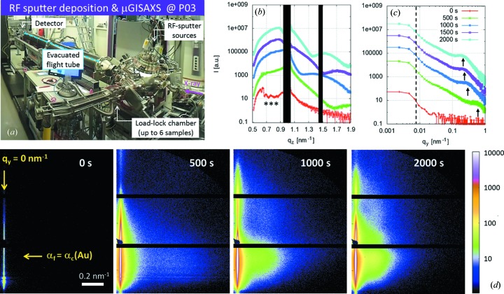
Figure 8.
(a) Photograph of the RF sputter deposition experiment set-up at MiNaXS. (b) Detector cut I[q z(t)] at q y = 0 nm−1. The black areas correspond to the shadow of the specular beamstop and to the gaps in between modules of the Pilatus 300k detector. The curves are shifted for clarity. Resonant diffuse scattering stemming from the thin polymer film is indicated by the stars (***). (c) Out-of-plane cut I[q y(t)] at αf = αc(Au). The curves are shifted for clarity. (d) µGISAXS data recorded at the MiNaXS beamline at an X-ray energy of 13.0 ± 0.1 keV, an incident angle of 0.435 ± 0.005°, a sample-to-detector distance of 4823 ± 2 mm and with an exposure time of 95 ms (exposure period 100 ms). The arrows indicate the positions of the detector (vertical arrow) and out-of-plane cuts (horizontal arrow) shown in (b) and (c), respectively. In this configuration the resolution limit is imposed by the beam divergence as indicated by the vertical dashed line at q y = 0.005 ± 0.001 nm−1 in (c).

