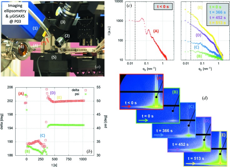
Figure 9.
Combined µGISAXS and imaging ellipsometry at MiNaXS. (a) Photograph of the experiment set-up (sample environment): (*) needle, highlighted by the red line; (1) ellipsometer laser arm; (2) ellipsometer detector arm; (3) optical microscope; (4) diode beamstop; (5) sample stage; (6) flight tube entrance window. (b) Ellipsometer data: delta (Δ) and psi (Ψ) as a function of time. (c) Out-of-plane cuts I[q y(t)] at αf = αc(Si) as indicated by the arrows in the two-dimensional GISAXS data shown in (d): (A) t < 0 s, (B) t = 0 s, (C) t = 366 s, (D) t = 452 s and (E) t = 513 s. The time that a droplet of the gold nanoparticle solution was deposited onto the polymer template defines t = 0 s.

