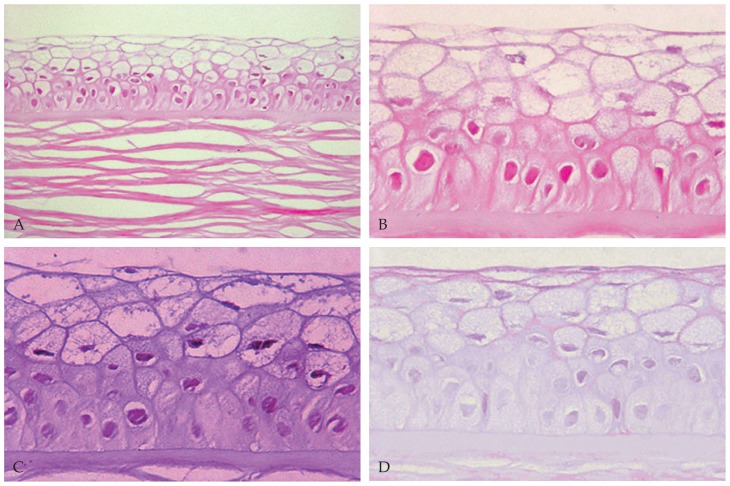
Figure 2.
Abnormal corneal epithelium with cellular swelling and intracytoplasmic cyst-like inclusions (A & B) on Hematoxylin & Eosin staining (A: magnification × 400, B: magnification ×1000). Note the presence of moderate amounts of periodic acid-Schiff-positive (C) and diastase-sensitive (D) material within the abnormal epithelial cells (magnification ×1000).

