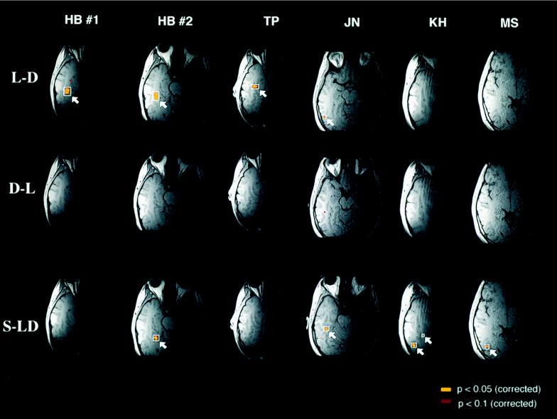
Figure 1.
Significant differences in the blood-oxygenation level-dependent (BOLD) MRI signal during passive viewing of letters vs. digits (L-D), digits vs. letters (D-L), and shapes vs. letters and digits (S-LD) (T.A.P., M. Stallcup, G. K. Aguirre, D. Alsop, M. D’Esposito, J. Detre & M.A.F., unpublished work). For each of the three comparisons in each of the six sessions, the single horizontal brain slice that showed the most significantly activated voxels for that comparison is shown. Voxels in yellow were significant at the P < 0.05 level after correcting for all the voxels as well as for the three planned comparisons. Voxels in red were significant at the P < 0.1 level, corrected. The left hemisphere appears on the left and the right hemispheres on the right.

