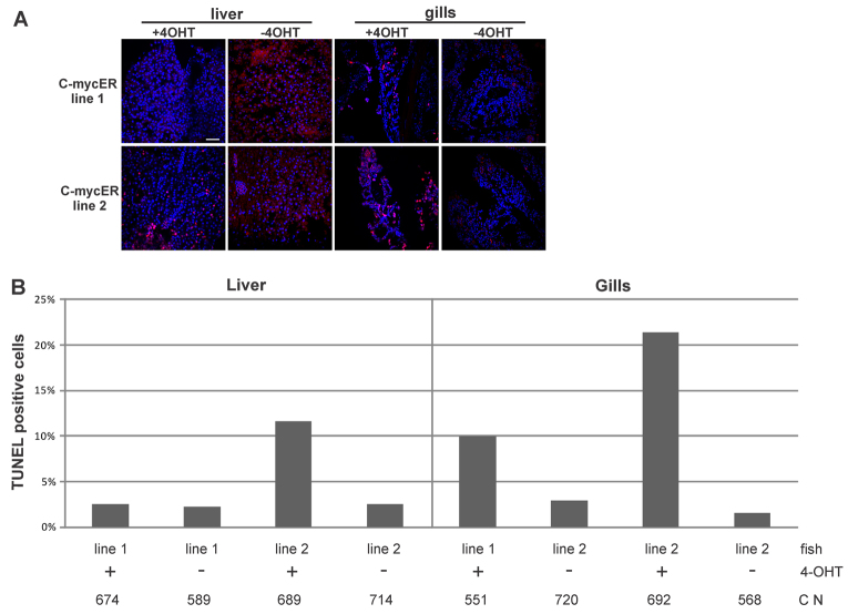
Fig. 6.
Induction of apoptosis after myc17 activation in vivo. (A) Detection of apoptosis by TUNEL assays in liver and gill sections from adult fish. Fish were treated for 24 hours with 4-OHT. DNA fragmentation is visible as red spots colocalizing with nuclei, which are stained with Hoechst. Scale bar: 10 μm. (B) Quantification of the percentage of apoptotic cells in liver (left panel) and gills (right panel) of both myc17ER lines in the presence or absence of 4-OHT. C N indicates total cell number for each assay.

