Fig. 2.
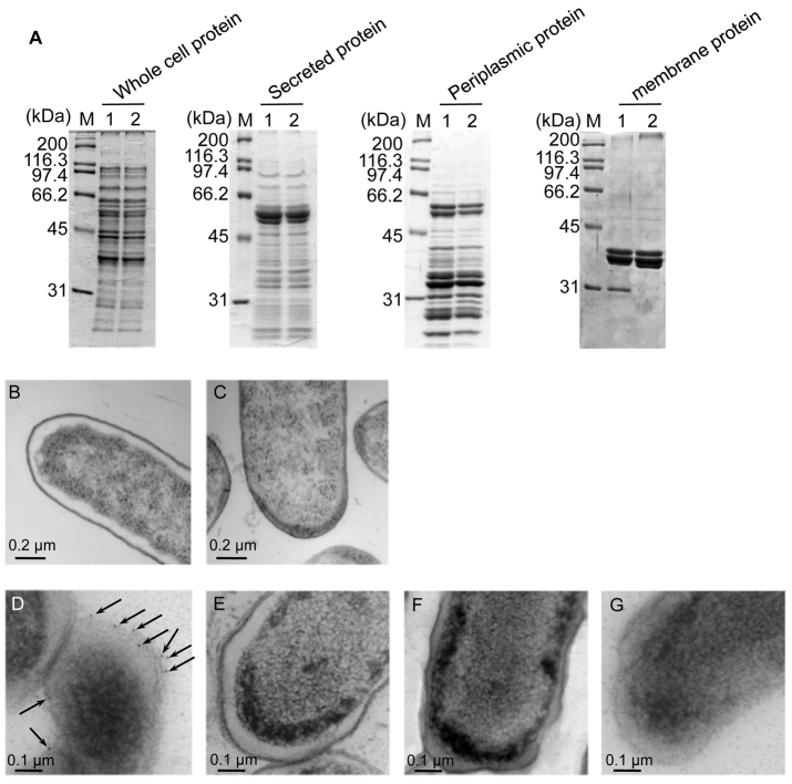
Regulation of OmpA localization by Stn. (A) SDS-PAGE of prepared protein fractions. Proteins were loaded on 10% gel in each lane as follows: 10 μg of whole cell protein fraction, 5 μg of secreted protein fraction, 10 μg of periplasmic protein fraction and 5 μg of total membrane protein fraction. Gel was stained with Coomassie Brilliant Blue R-250. M, protein marker; lane 1, wild type; lane 2, Δstn. (B,C) Transmission electron microscopic image of wild-type (B) and Δstn mutant (C) cells in ultrathin sections (magnification: 12,000×). Bacteria were cultured in LB medium at 37°C for 16 hours. (D–G) Immunogold labeling of OmpA using anti-OmpA antibody (magnification: 20,000×). Bacteria were cultured on LB agar plates at 37°C for 16 hours. D, wild-type strain; E, Δstn; F, ΔompA; G, wild type using normal mouse serum for control reaction. Arrows indicate the gold particles.

