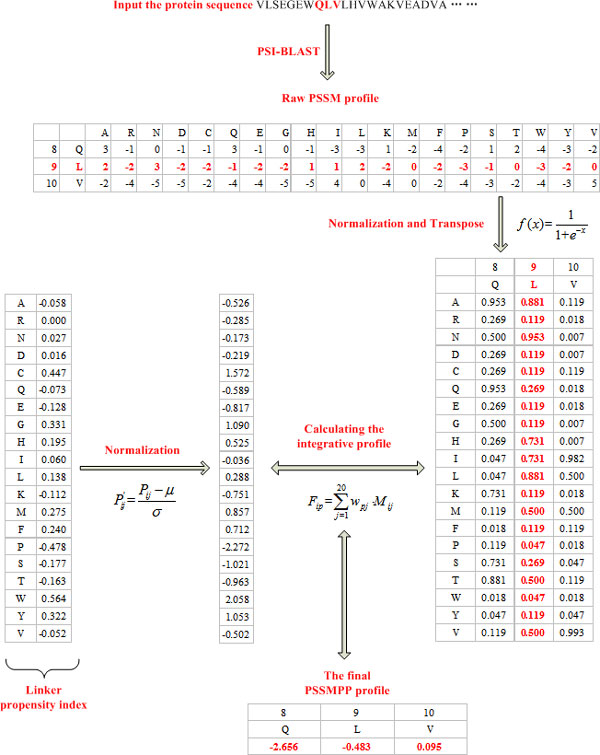
Figure 2.
Flowchart of generating the PSSMPP profile. Given a heme binding protein sequence (PDB id: 1A6M; Chain: A), a window size of 3 is set for a simple illustration. The central residue is 9 L (residue number in the sequence; residue name), with its two neighbouring residues on both sides (8 Q and 10 V).

