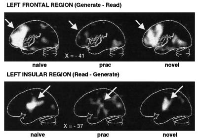
Figure 3.
PET difference (subtraction) images showing areas of increased (Upper images) and decreased (Lower images) blood flow when verb generation (under Naive, Practiced, and Novel conditions) is compared with reading. During naive (Left images) and novel (Right images) verb generation, increased blood flow in left frontal cortex was found compared with simple reading, whereas decreased blood flow was observed in left insular cortex. The Center images show that blood flow in these areas changed to a level almost identical to that found during simple reading after the verb generation was practiced. A linear gray scale is used with white representing maximal activation and black, minimal activation. The brain outlines were traced from the stereotaxic atlas of Talairach and Tournoux (20) and represent sagittal sections with their x-axis (left–right axis, left being negative) positions in millimeters noted.

