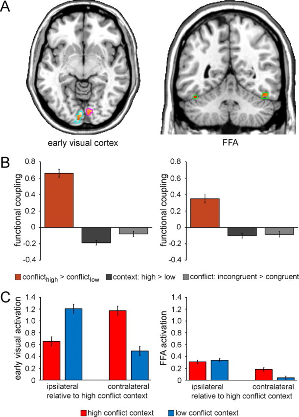
Figure 4.

Context-specific improvements in interference resolution are mediated by top-down control. A, Functional coupling with the right mSPL as a function of context-specific variation in conflict processing (interferencehigh-conflict context > interferencelow-conflict context) as revealed by PPI analysis is shown for voxels in the independently localized early visual cortex [left panel; left hemisphere (light blue outlines): x −2, y −86, z −12; 290 voxels; right hemisphere (magenta outlines): x 10, y −80, z −8; 75 voxels] and FFA ROIs (right panel; green outlines; left hemisphere: x −44, y −46, z −26; 35 voxels; right hemisphere: x 48, y −56, z −22; 53 voxels), displayed at p < 0.05 (small volume correction) on axial (z −7) and coronal (y −50) slices of an individual brain in MNI space. B, Greater functional coupling (β estimates ± SEM) between the mSPL and bilateral early visual cortex (left panel) and FFA (right panel) as a function of context-specific variation in conflict processing (orange) relative to that as a function general context representation (dark gray) and conflict processing (light gray) as revealed by PPI analysis (one-way ANOVAs: both F(1,24) > 29.1, both p < 0.0001). C, Mean activation (β estimates ± SEM) in the early visual cortex (left panel) and FFA (right panel) ROIs is plotted as a function of the laterality of the anatomical hemisphere (relative to the high-conflict location/visual hemifield: ipsilateral vs contralateral) and context (high conflict vs low conflict). Note that activation is not plotted relative to stimulus presentation per se given the counterbalancing of the contextual conflict frequency manipulation across participants.
