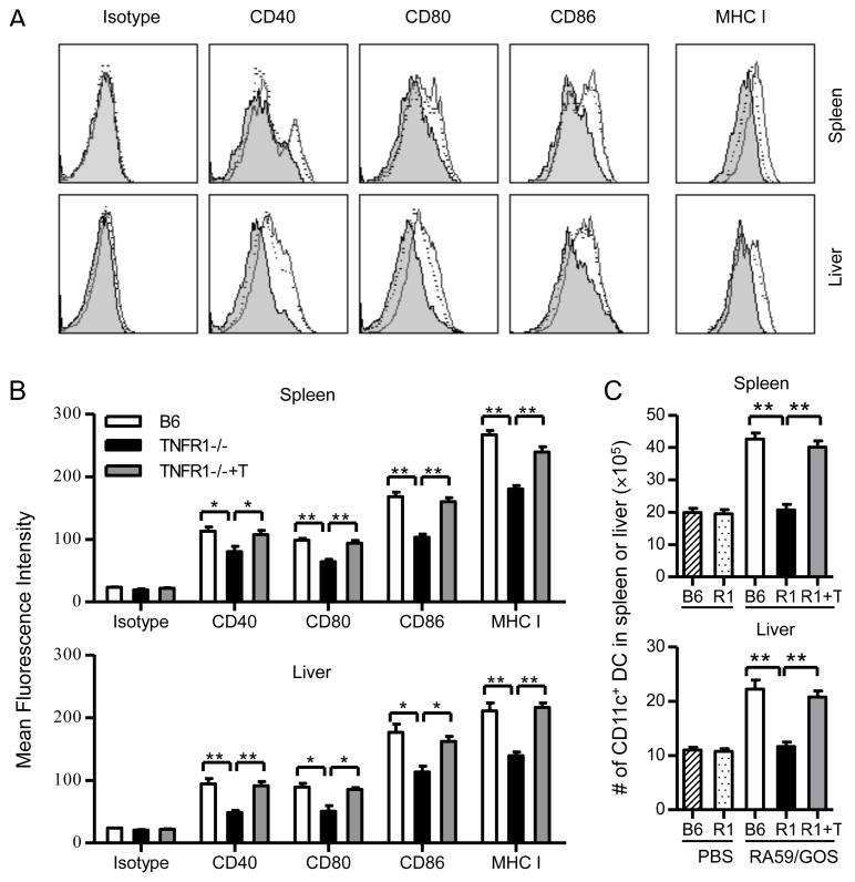
FIGURE 4. DC maturation and mobilization is impaired in TNFR1-deficient mice and the impaired DC response is restored by adoptive transfer of TNFR1-positive T cells.
B6 mice, TNFR1−/− mice and TNFR1−/− mice that were transferred with purified CD3+ T cells (TNFR1−/−+T or R1+T) one day earlier were inoculated with 1 × 10⁶ pfu of RA59/GOS virus or the same volume of PBS. Three days later, cells from spleen and liver were enumerated and analyzed for CD11c plus CD40, CD80, CD86 or MHC class I. (A) Comparison of CD40, CD80, CD86 and MHC I expression by CD11c+ cells from B6 mice (histograms with solid lines), TNFR1−/− mice (shaded histograms), and TNFR1−/− mice injected with T cells (histograms with dotted lines). (B) Comparison of mean fluorescence intensity (MFI) of CD40, CD80, CD86, and MHC I by CD11c+ cells from the spleen (upper panel) or liver (lower panel) from B6 mice, TNFR1−/− mice, and TNFR1−/− mice injected with T cells. (C) Comparison of the total numbers of CD11c+ cells in the spleen (upper panel) and liver (lower panel) of B6 mice, TNFR1−/− mice (R1), and TNFR1−/− mice injected with T cells (R1+T). * p< 0.05; ** p< 0.01.

