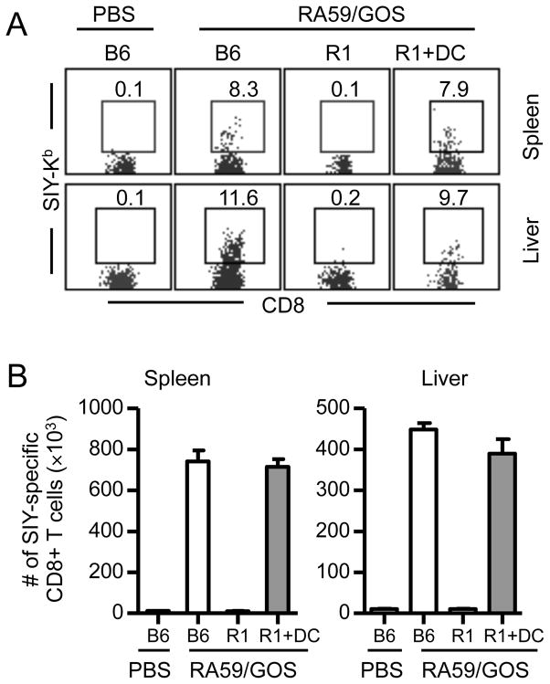
FIGURE 5. Rescue of CD8 T cell response in TNFR1-deficient mice by adoptive transfer of TNFR1-expressing DCs.
Bone marrow cells from B6 mice were cultured in the presence of GM-CSF for 7 days. DCs (75% CD11c+) were injected intravenously into TNFR1−/− mice (R1+DC, 8 × 10⁵ per recipient). One day later, mice were infected with 1 × 10⁶ pfu of RA59/GOS virus and the frequency and the number of SIY-specific CD8 T cells were analyzed in the spleen and liver at 7 dpi as in Fig. 2. (A) Representative SIY-Kb versus CD8 staining profiles of CD3+ CD8+ cells from spleen and liver. (B) Comparison of mean ± SD of SIY-Kb+ CD8+ cells in the spleen and liver of 4 mice per group. Data shown are from one of two independent experiments.

