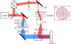
Fig. 1.
Synthetic aperture microscopy setup. Laser: He-Ne laser; GM: galvanometer scanning mirror, L1: lens; BF: back focal plane of condenser lens; C: condenser lens; OL: objective lens; : tube lens, ; AOMs: acousto-optic modulators; SF: spatial filter system. Frequency shifted reference laser beam is shown in blue. Inset: spiral trajectory of the focused spot at the BF.

