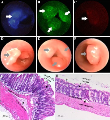
Fig. 4.
Individually targeted in vivo images. Colonic adenomas (arrows) are localized on wide-field fluorescence images collected with the multispectral endoscope using separately administered peptides (a) KCCFPAQ-DEAC, (b) AKPGYLS-TAMRA, and (c) LTTHYKL-CF633. The corresponding white light images are shown in (d–f). Routine histology (H&E) of (g) adenoma shows features similar to those seen in sporadic human adenomas, including enlarged nuclei, hyperchromaticity, and distorted crypt architecture, and (h) normal appearing adjacent colonic mucosa, scale bar 100 μM

