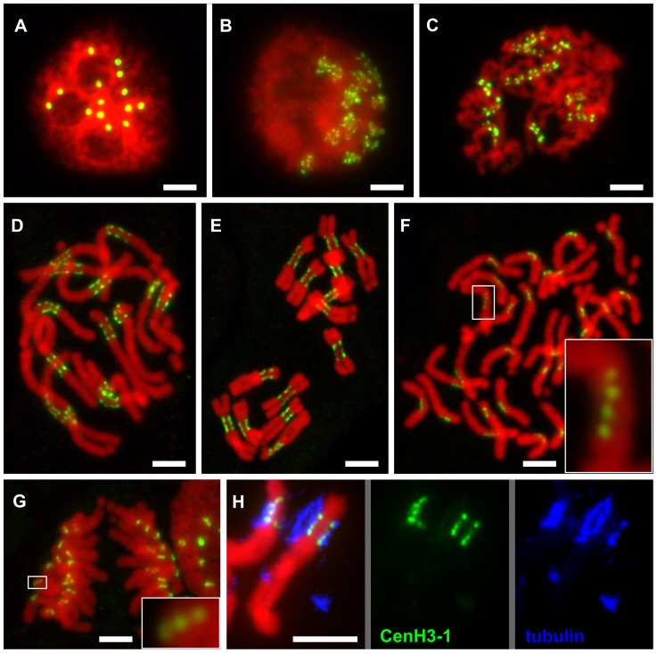
Figure 2. Organization of CenH3-containing domains during the cell cycle.
A: Number of CenH3-containing domains in interphase nucleus corresponds to the number of chromosomes in diploid cells (2n = 14). B–D: Mitotic chromosomes at the early prophase (B), prophase (C) and prometaphase (D) show multiple domains containing the CenH3 which are clearly separated with chromatin segments lacking the CenH3. E–G: The separation of CenH3-containing domains becomes less apparent with the progress of chromatin condensation: metaphase (E), spread of single-chromatid anaphase chromosomes (F), anaphase (G). However, the multiple domain structure can be observed in some cases even in the highly condensed anaphase chromosomes (detail windows in F and G). H: Simultaneous detection of CenH3-1 (green) and tubulin (blue) revealed that microtubules are attached to all CenH3 containing domains as shown for chromosome 3. All chromosomes were counterstained with DAPI (pseudocolored in red). Bar = 5 µm.

