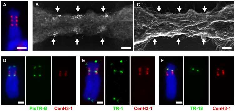
Figure 4. Organization and DNA sequence composition of CenH3-containing domains in chromosome 3.
A–B: Primary constriction of chromosome 3 contains three functional centromere domains as defined by the presence of CenH3-1. Correlative fluorescence and scanning electron microscopy images of the same chromosome using FluoroNanogold showed that the three domains recognized with fluorescence (A, red signals) are composed of multiple foci from markers (bright spots) near the surface of the primary constriction (B, backscattered electron micrograph). C: Secondary electron micrograph image of the same chromosome. The primary constriction exhibits few chromomeres and a typical longitudinal orientation of fibrillar substructures, to which the CenH3 domains roughly correspond. The arrows mark the CenH3-1 containing regions. D–F: Detection of three different families of satellite DNA by FISH (green) combined with immunodection of CenH3-1 (red). Each of the functional centromere domains is composed of different family of satellite DNA; the domain closest to the long arm is composed of PisTR-B (D), the middle one of TR-1 (E) and the one closest to the short arm of TR-18 (F). Chromosomes were counterstained with DAPI (blue). Bar = 2 µm (A and D–F) or 0.2 µm (B–C).

