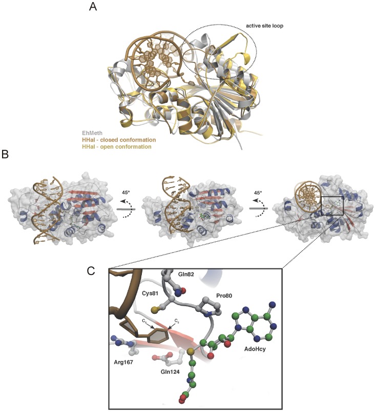
Figure 3. Model of the EhMeth-DNA complex.
(A) Superposition of EhMeth (grey) and M.HhaI in closed conformation (brown) and open conformation (yellow). The superposition clearly illustrates that the active site loop of EhMeth adopts a conformation between the open and the closed conformation of M.HhaI. (B) Due to the high structural homology between EhMeth and M.HhaI a DNA-binding model could be derived from superposition with the substrate bound M.HhaI-structure. The M.HhaI-DNA neatly fits into the putative DNA-binding site of EhMeth. (C) The close-up of the active site illustrates that the flipped out cytosine is in a conformation that would allow for methyl-group transfer further supporting, that EhMeth follows the same reaction mechanism as M.HhaI and DNMT2.

