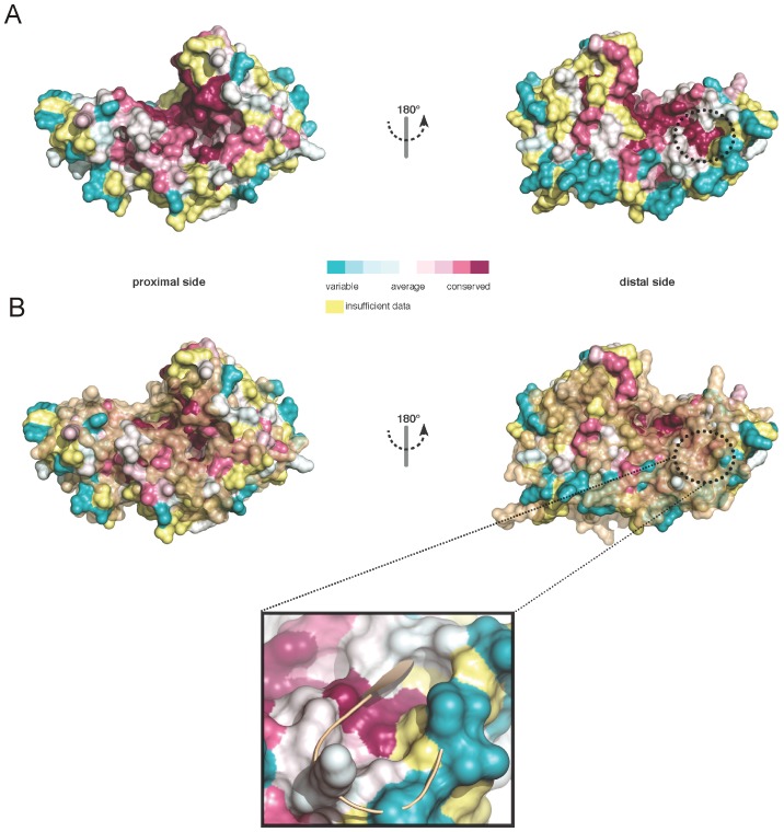
Figure 4. Conservation among DNMT2 enzymes.
(A) Conserved residues among DNMT2 MTases were mapped to the surface of EhMeth. Purple color displays high conservation, cyan color displays high variance. Highly conserved areas can be seen in the active site, the putative DNA/tRNA binding site as well as in a highly acidic pocket on the distal site of EhMeth. (B) A superposition of the conserved areas with the surface of M.HhaI (closed conformation, brown) shows that only minor divergence can be observed at the proximal site while in particular the acidic pocket on the distal side is covered – that is not existent in M.HhaI. The close-up illustrates that the acidic pocket is occupied by strand β10.

