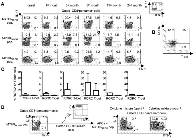
Figure 6. Functional flexibility of fresh IL-17–producing CD8+ T cells specific to apoptotic epitopes.
(A) PBMCs from three independent patients with acute HCV infection were stained with mAb to CD8 and pentamers complexed with the indicated apoptotic peptide. Cells were stimulated with the relevant soluble peptide plus anti-CD28 mAb for 6 h and then processed for the detection of IL-17 and IFN-γ by ICS assay with the relevant mAbs. Counterplot analyses are gated on CD8+pentamer+ cells and show percentages of cytokine-producing cells. The percentage of cells is reported in each quadrant. The small histograms show ICS analyses of representative cytokine (IL-17, or IFN-γ) production without antigenic stimulation by gated CD8+pentamer+ cells. (B) Representative flow cytometry analysis in which cells were stimulated as previously described and processed for the detection of IL-17, IFN-γ, RORC, and T-bet by ICS assay with the relevant mAbs. Analysis gated on CD8+pentamer+IL-17+ IFN-γ+ cells (signed by the star symbol in the panel A) show percentages of RORC- and Tbet-expressing cells. The percentage of cells is reported in each quadrant. (C) Mean percentages of RORC+ or T-bet+ cells detected in all the timely followed CD8+pentamer+ cells producing IL-17 or IFN-γ upon antigen stimulation. (D)IL-17–producing CD8+ T cells specific to apoptotic epitopes efficiently convert to IFN-γ producing cells in vitro. One representative of three experiments in which PBMCs from patients with acute HCV infection were stained with mAb to CD8 and pentamers complexed with the indicated apoptotic peptide, stimulated with the relevant soluble peptide plus anti-CD28 mAb for 6 h, and processed for the detection of IL-17 and IFN-γ by ICS assay with the relevant mAbs. Counterplot analyses are gated on CD8+pentamer+ cells and show percentages of cytokine-producing cells. The percentage of cells is reported in each quadrant. Cells producing IL-17 were sorted by using anti-CCR6 and anti-CCR4 mAbs and FACSAria processing (the purity of IL-17+ cell population was evaluated by stimulating CCR6+CCR4+ cells with anti-CD3 and anti-CD28 mAbs). CCR6+CCR4+ cells were re-stimulated with the same apoptotic epitope and autologous APCs in the presence or absence of either the cytokine mixture type-17 (IL-1β, IL-6, IL-23 and TGF-β) polarizing toward the type-17 cell phenotype or the cytokine mixture type-1 (IFN-γ and IL-12) polarizing toward the type-1 cell phenotype. After 10–12 d of culture in IL-2 conditioned medium, cells were stained with pentamers expressing MYH9741–749 peptide, further antigen-stimulated as previously described, and tested for their capacity to produce IL-17 and IFN-γ by ICS assay.

