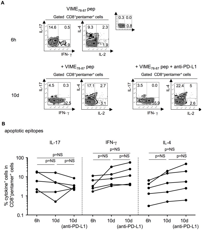
Figure 7. Tuning of apoptotic epitope-specific CD8+ T cells by PD-1.
(A) Representative experiment in which PBMCs from a patient with acute HCV infection were stained with mAb to CD8 and the pentamer complexed with the indicated apoptotic epitope. Cells were then stimulated with the soluble form of the indicated peptide and anti-CD28 mAb in the presence or absence of a blocking antibody to PD-L1 or an isotype control. After 6 h and 10 d of culture, cells were processed for the detection of the indicated cytokines by ICS assay with the relevant mAbs. (B) Cumulative experiments, performed as described in (a), showing the percentages of cells producing the indicated cytokines in CD8+pentamer+ cells, evaluated at different time points from the stimulation with the apoptotic peptides in the presence or absence of mAb to PD-L1. NS = not significant.

