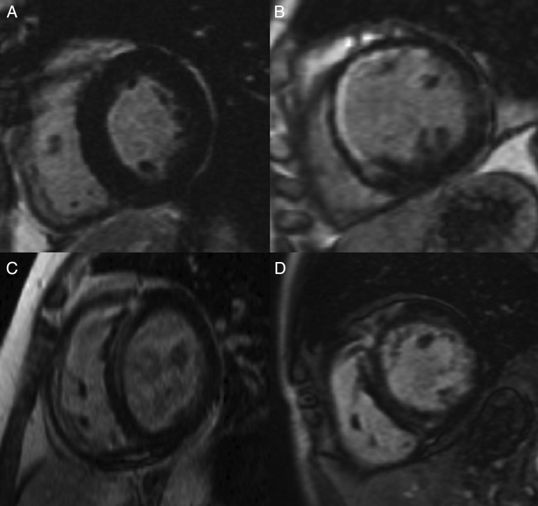
Figure 1:
Examples of the different patterns of LGE on steady-state free procession images of the left ventricle in end-diastole. (A) Short axis view of the left ventricle in a patient with no LGE. (B) Infarct pattern of LGE with subendocardial enhancement of the antero-septum and anterior wall. (C and D) Linear mid-wall fibrosis predominantly affecting the LV septum (mid-wall LGE). The patient in (C) went on to develop heart block perioperatively.

