Figure 4:

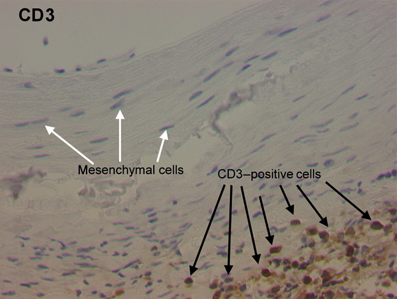
The representative immunohistochemical staining of an elastase-treated animal (CD3). Note the T-lymphocyte invasion into the adventitia of the vessel wall.

The representative immunohistochemical staining of an elastase-treated animal (CD3). Note the T-lymphocyte invasion into the adventitia of the vessel wall.