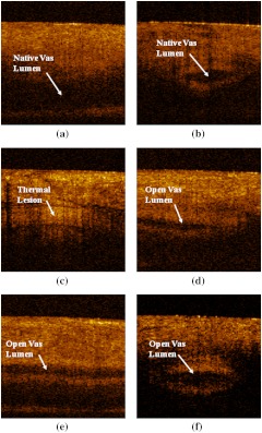
Fig. 3.
(a) and (b) Longitudinal and cross-sectional OCT images of the native canine vas acquired while the vas and scrotal skin was compressed inside the vas ring clamp prior to the procedure. (c) Longitudinal OCT image of the thermally coagulated canine vas acquired while the vas and scrotal skin were compressed inside the vas ring clamp after the procedure. (d) Longitudinal OCT image of the thermally coagulated canine vas acquired ex vivo after the vas tissue was harvested; (e) and (f) longitudinal and cross-sectional OCT images of the native canine vas acquired ex vivo after the tissue was harvested. All OCT images measure ().

