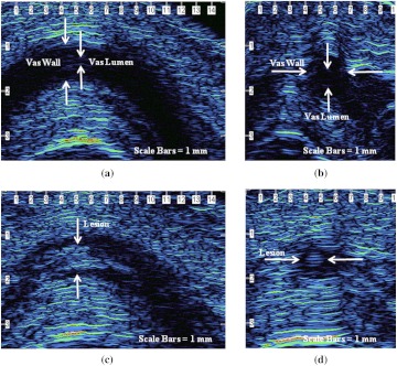
Fig. 4.
(a) and (b) Longitudinal and cross-sectional HFUS images of the native canine vas, acquired by manually isolating the vas within the scrotal skin fold prior to placement of the vas ring clamp. The vas wall and lumen can be identified. (c) and (d) Longitudinal and cross-sectional HFUS images of the thermally coagulated canine vas acquired by manually isolating the vas within the scrotal skin fold after removal of the vas ring clamp. A thermal lesion encompassing both the vas wall and lumen is observed.

