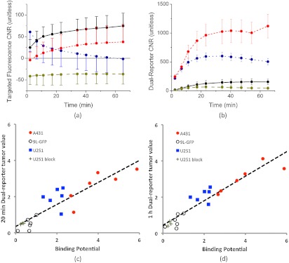
Fig. 4.
Targeted fluorescence uptake versus dual-reporter imaging within 1 h of reporter injection. The error targeted fluorescence uptake tumor contrast-to-noise ratio (CNR) within 1 h of reporter injection is presented in (a) for each tumor group (; ; ; ). The error tumor CNR determined by the dual-reporter approach for the same tumor groups as in (a) is presented in (b). The correlation between the average dual-reporter image value of the tumor in each mouse at 20 min and 1 h after reporter injection and the corresponding binding potential (an in vivo marker of receptor expression) are presented in (c) and (d), respectively. Data points from each tumor line are color-coded to match the data in (a) and (b). The slope of the 20-min correlation was (, ), and the 1-h correlation was (, ).

