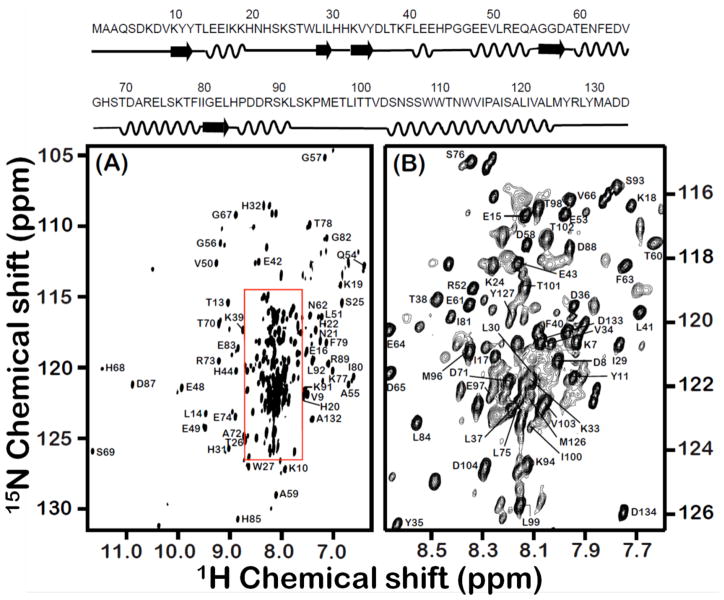
Figure 1. Backbone amide resonance assignment for the 16.7-kDa rabbit cytb5 incorporated in DPC micelles.
A full 2D 1H-15N TROSY-HSQC spectrum of U-13C, 15N, 2H labeled full-length cytb5 in DPC micelles along with resonance assignments is shown in panel A while panel B represents expanded region of the box shown in panel A. The amino acid sequence with its secondary structure is shown at the top.

