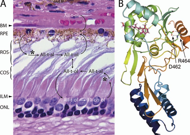
Figure 2.
Overview of the visual cycle, and structure of an IRBP module. A) The diagram is overlaid on a hematoxylin & eosin stained paraffin section of human retina (near the fovea). Interphotoreceptor matrix (IPM) glyconjugates are largely responsible for the basophilic outer segment staining. The apparent spaces among the outer segments is a histological processing artifact. The larger cone nuclei are located near the external limiting membrane (ELM, arrowhead). Bruch’s membrane (BM, arrow head); outer nuclear layer (ONL). 11-cis retinal (11-cis-al) is photoisomerized (*) to its 11-cis isomer, which is reduced to all-trans retinol (All-t-ol). The All-t-ol is then released from rod and cone outer segments (ROS and COS respectively) into the IPM. B) Ribbon representation of the module II Xenopus IRBP structure docked with all-t-ol (magenta).The N- and C-terminal regions are shown in blue and red respectively. A salt bridge (dotted lines) extends between the carboxamide side group of D462, and guanidinium group of R464. These highly conserved residues are shown in stick representation (oxygen, red; nitrogen, blue). The corresponding aspartic acid of human IRBP is replaced by asparagine in a form of autosomal recessive retinitis pigmentosa. It is possible that this substitution (D1080N), by abolishing the conserved salt bridge, destabilizes the nearby retinoid-binding site. The structure shown in this panel is adapted from Hollander et al. Invest. Ophthal. Vis. Sci. 50:1864–72, 2009 with permission from the Association for Research in Vision and Ophthalmology.

