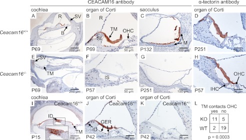
FIGURE 5.
Expression of CEACAM16 in the cochlea and vestibular organ. Expression of CEACAM16 was analyzed in the cochleae and vestibular organ of Ceacam16+/+ (A–C, I, and J) and Ceacam16−/− mice (E–G) by immunohistology using the biotinylated monoclonal anti-human CEACAM16 antibody 9D5, which cross-reacts with murine CEACAM16. CEACAM16 (brown stain) was observed in the acellular TM (A, B, I, and J) and in the extracellular matrix covering the macula cells of the saccule (C) in wild-type but not in Ceacam16−/− mice. Furthermore, staining for CEACAM16 was observed in interdental cells in the helicotrema region of P15 wild-type cochleae (I) and also in adult Ceacam16+/− mice (J). No staining was seen when only horseradish peroxidase-coupled streptavidin was added (K). Comparable staining for α-tectorin was found in the TM in both Ceacam16+/+ (D) and Ceacam16−/− mice (H). The age of the mice (days post partum) is indicated at the lower left corners. Note that the TM of Ceacam16−/− mice is significantly more often in contact with the OHCs (E, F, and H), whereas it is either detached or more retracted in wild-type mice (B, D, I, and J). No difference was observed when different regions of the cochlea (base, medal region, or helicotrema-proximal) were analyzed. Five wild-type and four Ceacam16−/− mice, representing eight and seven cochleae, respectively, were evaluated; p = 0.0003, Fisher's exact test (L). B, basilar membrane; GER, greater epithelial ridge; ID, interdental cells; IS, inner sulcus; M, macula; R, Reissner's membrane; SV, stria vascularis; SL, spiral limbus; TM, tectorial membrane.

