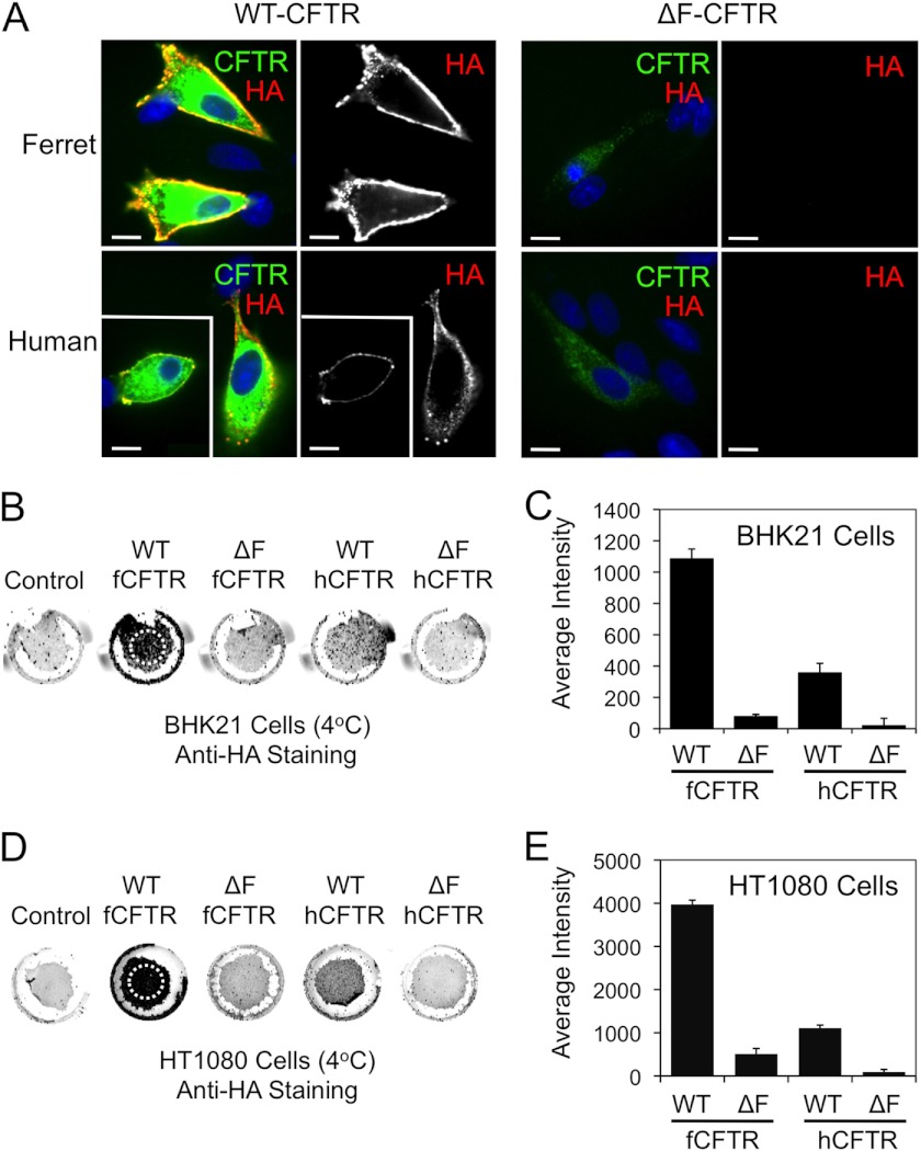
FIGURE 5.
Ferret and human ΔF508-CFTR lack significant cell surface expression relative to the wild-type counterparts. A, BHK21 cells grown on coverslips were transfected with the indicated plasmid expression constructs. At 48 h post-transfection, the cells were brought to 4 °C, and a mouse anti-HA antibody was applied for 1 h to stain surface HA-tagged CFTR (red). The unbound antibody was washed away, and the cells were then fixed and permeabilized. The cells were then stained for total CFTR using a rat anti-CFTR antibody (green) and DAPI to mark nuclei (blue). Immunofluorescent images of ferret and human WT-CFTR (left panels) and ΔF508-CFTR (right panels) are shown with the single anti-HA channel shown to the right of the merged color images. Boxed insets show a second example to the larger image. Scale bars represent 10 μm. B–E, surface On Cell Western blots were performed by binding an anti-HA antibody at 4 °C to cell surface CFTR on live BHK21 (B and C) and HT1080 (D and E) cells that were transfected with the indicated constructs as in A and a non-HA tagged CFTR control construct. Excess anti-HA antibody was washed away at 4 °C followed by fixation and staining with an IR secondary antibody. Wells were then imaged using an Odyssey IR scanner. The resulting example images (B and D) were quantified using ImageJ software to give the data shown in (C and E). Only regions containing cells, as indicated by the hashed circles on the WT-fCFTR samples (B and D) were evaluated for the average intensity of staining (which is independent of the area of the field quantified). This was necessary because variable sloughing of cells occurred during the assay washings. Data in all graphs represent the control background subtracted mean ± S.E. with n = 16 from 4 independent experiments. All comparisons with the exception of human ΔF508-CFTR versus ferret ΔF508-CFTR were statistically significant as determined by the Student's t test (p < 0.001). Human ΔF508-CFTR versus ferret ΔF508-CFTR was not significant for BHK21 cells but was significant for HT1080 (p < 0.008). With the exception of ferret ΔF508-CFTR in HT1080 cells, all other ΔF508-CFTR analyses were not significantly above control background levels of cells transfected with a non-HA-tagged version of CFTR.

