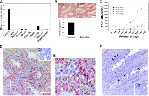
FIGURE 1.
Expression profiles of miR-449 cluster and miR-34 family in mice. A, qPCR analyses of expression levels of miR-449 in 11 tissues collected from adult male mice. B, germ cell depletion through busulfan treatment followed by qPCR detection of miR-449. miR-449 was abundant in control (untreated wild type) testes, whereas miR-449 was undetectable in busulfan-treated (germ cell-depleted) testes, suggesting that miR-449 is exclusively expressed in germ cells. Scale bar, 50 μm. C, qPCR analyses of expression levels of miR-449 and members of the miR-34 family during postnatal testicular development. D, localization of miR-449 in the mouse testis using in situ hybridization. Hybridization signals are in red (Texas Red), and the nuclei were counterstained with hematoxylin (blue). The inset is a control. Scale bar, 70 μm. E, an enlarged image of the field framed be the dashed box in D. Specific miR-449a hybridization signals were mainly detectable in the cytoplasm of spermatocytes and spermatids (arrowheads), whereas no signals were fund in somatic cell types and spermatogonia (arrows). Scale bar, 50 μm. Ctr, control. F, localization of miR-34a in the murine testis by in situ hybridization. Hybridization signals (red) were only detected in spermatogonia. Cell nuclei (blue) were counterstained with hematoxylin (blue). The inset is a control. Scale bar, 50 μm. Data are represented as mean ± S.E. (n = 3).

