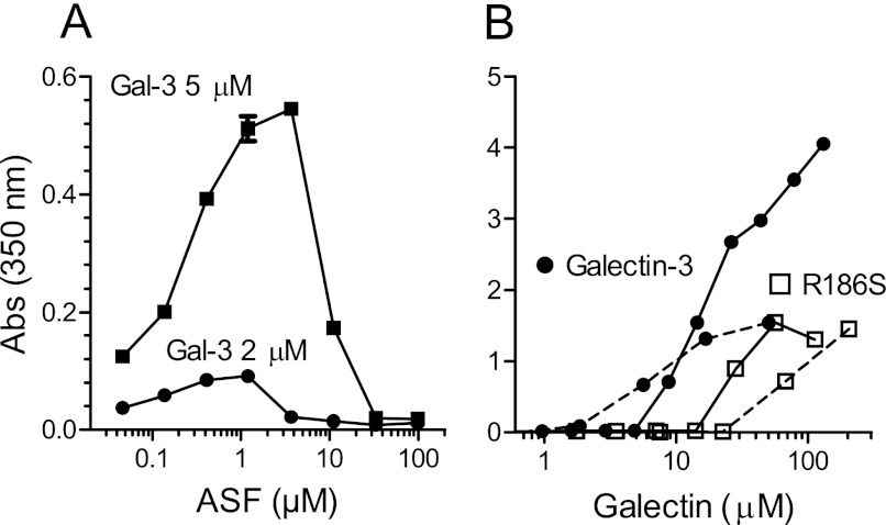
FIGURE 3.
Detection of galectin-3-ASF precipitates. A, fixed concentrations of galectin-3 (2 and 5 μm) were mixed with a range of concentrations of ASF. B, fixed concentrations of ASF (0.8 μm (broken lines) or 20 μm (unbroken lines) were mixed with a range of concentrations of galectin-3. Absorbance (Abs) was measured at 350 nm (y axes), at room temperature within 10 min. Background (∼0.2), mainly due to the plate, was subtracted. Absorbance due to the components of the samples measured separately was <0.02.

