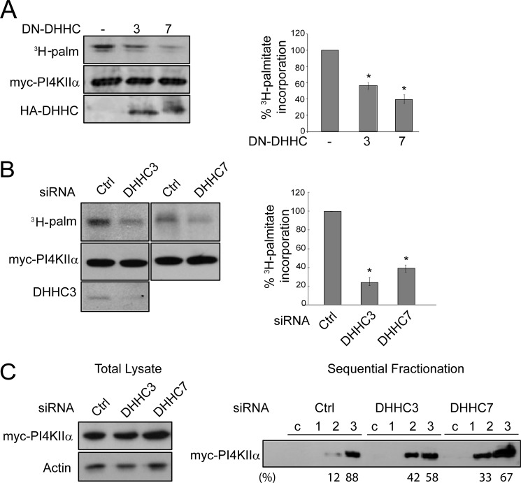
FIGURE 2.
Effect of DN-DHHC3 or DN-DHHC7 inhibition or silencing on PI4KIIα palmitoylation. Representative Western blots and fluorography are shown. Palmitoylation was quantified by ImageQuant 5.2 software and is expressed as a percentage of the control. Values are means ± S.E. (n = 3). *, p < 0.01. A, overexpression of DN-DHHC3 or DN-DHHC7. COS-7 cells were cotransfected with Myc-PI4KIIα and either DN-DHHC3 or DN-DHHC7 and labeled with [3H]palmitate (3H-palm). Myc-PI4KIIα was immunoprecipitated, resolved by SDS-PAGE, and analyzed by fluorography and Western blotting. Left, Western blot and fluorography of a representative experiment; right, percentage 3H palmitoylation. B, DHHC3 or DHHC7 RNAi. HeLa cells were transfected with DHHC or control (Ctrl) siRNA for 48 h, followed by Myc-PI4KIIα transfection for another 16 h, and then labeled with [3H]palmitate for 4 h. Endogenous DHHC3 was detected with anti-DHHC3 antibody in Western blots. C, DHHC RNAi effect on PI4KIIα membrane extractability. Cells were homogenized, and the post-nuclear supernatant (lysate) was analyzed by Western blotting (left). The lysate was centrifuged at 200,000 × g to obtain the cytosolic fraction (c) and membrane pellet. Membranes were extracted sequentially with 1 m NaCl, 0.1 m Na2CO3, and 1% Triton-X100 (fractions 1–3, respectively) (right). Equivalent amounts of each fraction were analyzed by immunoblotting with anti-Myc antibody. The percentage of total PI4KIIα in each fraction is indicated below the Western blot.

