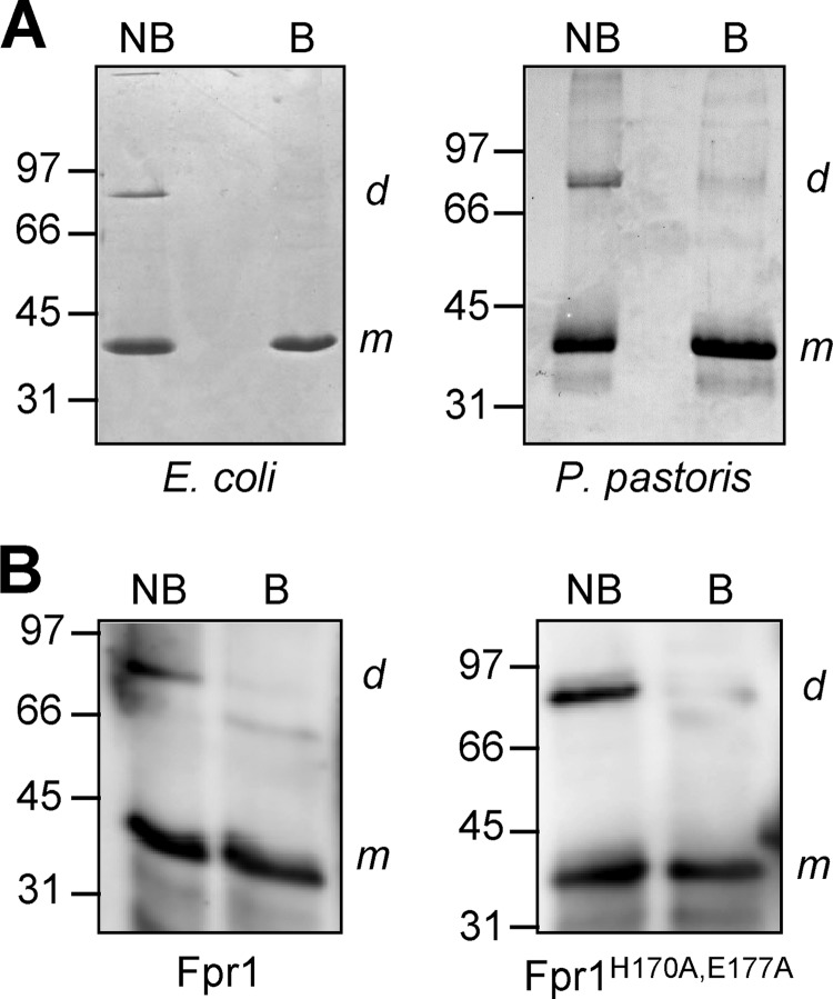
FIGURE 3.
Fpr1 forms a dimer in solution. A, 10-μg recombinant Fpr1 protein obtained from E. coli or P. pastoris was subjected to SDS-PAGE either without (NB) or with 5 min of boiling (B) prior to loading. Proteins were stained with Coomassie Blue. B, Western blot analysis of recombinant Fpr1 or FprH170A,E177A protein obtained from E. coli, subjected to SDS-PAGE either without (NB) or with 5 min of boiling (B) prior to loading and hybridized with α-Fpr1 antiserum. m and d indicate positions of hybridizing bands corresponding to the monomeric and dimeric form of Fpr1, respectively. Relative positions of molecular weight markers (in kDa) are indicated at the left.

