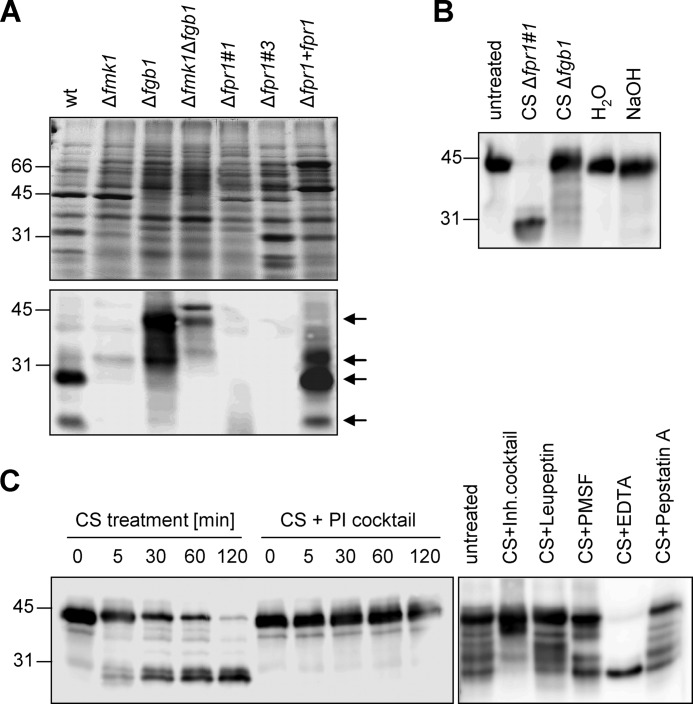
FIGURE 5.
Fpr1 is processed proteolytically. A, Western blot analysis of culture supernatants of different F. oxysporum strains with α-Fpr1 antiserum. The arrows point to hybridizing bands of different molecular weight. B, immunoblot of Fpr1 protein incubated for 16 h in the presence of culture supernatant (CS) of the indicated fungal strains, water, or 0.2 N NaOH. C, immunoblot of recombinant Fpr1 protein incubated with culture supernatant of strain Δfpr1–1 for the indicated time periods (left panel) or for 16 h (right panel) in the absence or presence of a protease inhibitor (PI) cocktail or of the indicated protease inhibitors. Relative positions of molecular weight markers are indicated at the left.

