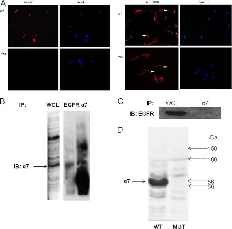
FIGURE 7.
Interaction between α7nAChR and EGFR. A, mouse sperm were smeared on a slide for immunocytochemical staining with anti-α7 or anti-EGFR antibodies as described under “Experimental Procedures.” The arrows indicate the acrosome. Larger magnification appears in the upper left corner of each photograph. Samples of 5 × 10⁷ mouse WT (B) or α7-null sperm (C) were washed in TBS and resuspended in homogenization buffer. The homogenate was then sonicated at 40 Hz, three times for 10 s each, and rotated at 4 °C for 30 min. The samples were immunoprecipitated (IP) and then precleared for 1 h at 4 °C, and antibodies, anti-EGFR (C) or anti-α7 (B and D), were added overnight. The next day, protein A/G was added for 5 h at 4 °C, and the samples were washed four times in TBS containing 0.1% Triton X-100. The washed beads were resuspended in sample buffer, and the sample was boiled for 5 min. The samples were separated in SDS-PAGE as described under “Experimental Procedures,” and the membrane was incubated with anti-α7 and goat anti-rabbit secondary antibody or anti-EGFR and goat anti-mouse secondary antibody. The data represent one experiment typical of three repetitions performed. D, proteins were extracted from WT or α7-null sperm with SDS lysis buffer as described under “Experimental Procedures.” The extracts were separated on SDS-PAGE and were transferred to nitrocellulose membrane that was incubated with anti-α7 as described under “Experimental Procedures.” The data represent one experiment typical of three repetitions performed. IB, immunoblot; WCL, whole cell lysate.

