FIGURE 8.
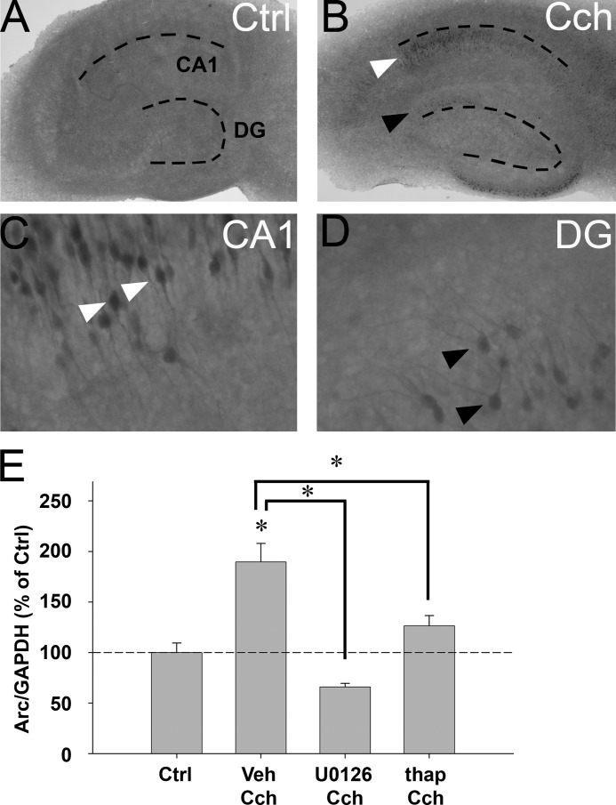
Cch induces Arc protein expression in rat organotypic hippocampal slice culture. A and B, immunohistochemistry reveals Arc protein expression in untreated slices (A, ctrl) and slices stimulated with Cch (50 μm) for 2 h (B, Cch). Arc-positive cells are visible in CA1 (white arrowhead) and dentate gyrus (DG, black arrowhead). Dotted lines indicate the DG- and CA1 cell layers. C and D, high magnification reveals Arc-positive CA1 pyramidal cells (C, white arrowheads) and dentate granule cells (D, black arrowheads). Note that Arc protein is found in dendrites, the somatic cytoplasm, and nucleus of granule cells and CA1 pyramidal cells. E, quantitative analysis of Western blots shows Arc protein expression in slices pretreated with U0126, thapsigargin (thapsi), or vehicle (veh) and then stimulated with Cch for 2 h. Arc protein levels are normalized to GAPDH and expressed in percent of Arc expression in untreated slices (Ctrl). Asterisks indicate a significant change relative to Ctrl and between groups (n = 4–5, p < 0.05).

