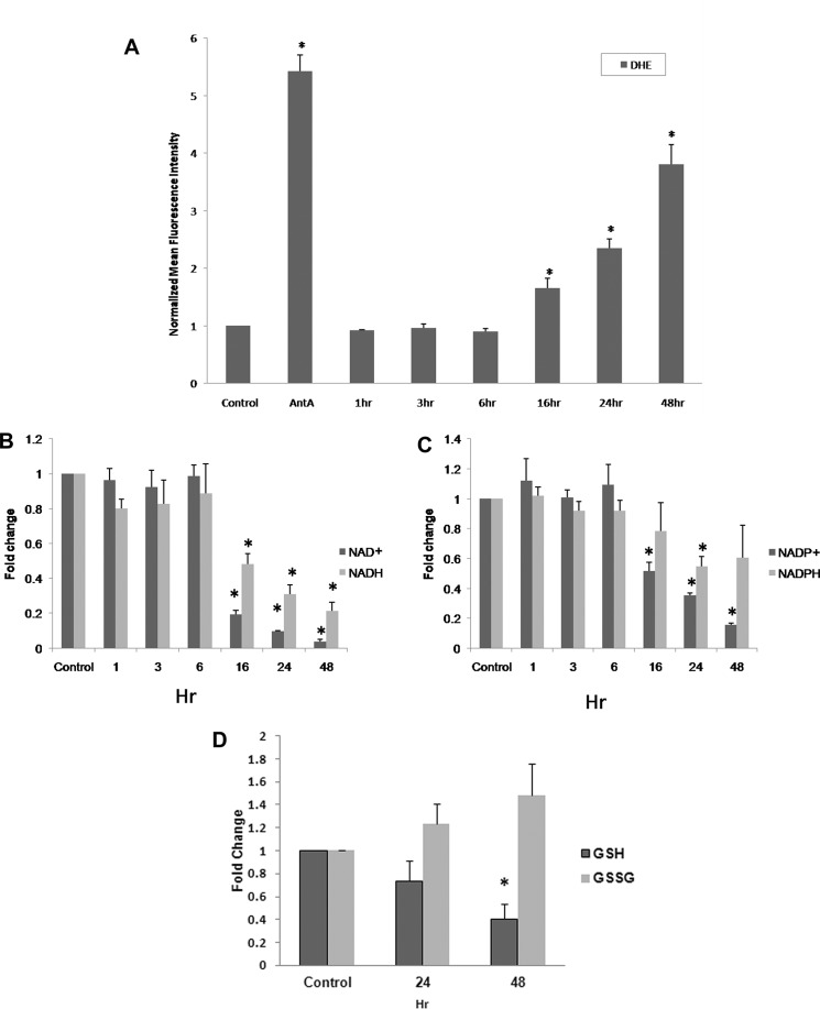
FIGURE 1.
A, increased steady state levels of superoxide demonstrated by increased DHE oxidation in human glioma cells, U251 with 5 nm GMX1778. Cells were plated for 24 h prior to GMX1778 treatment. Cells were exposed to GMX1778 for the times indicated prior to DHE treatment. Cells exposed to antimycin A (AntA, 10 μm) for 40 min during DHE treatment were used as a positive control. Error bars represent ± S.D. of three different separate experiments. (*, significantly different from DHE-only group, p < 0.05, n = 3.) B and C, fold changes in NAD+/NADH and NADP+/NADPH levels were measured after cells were treated with 5 nm GMX1778 for the indicated times. Fold change was calculated as the ratio of NAD+ or NADH and NADP+ or NADPH, respectively, per mg of protein normalized to control. Error bars represent ± S.D. of three different separate experiments. (*, significantly different from control group, p < 0.05, n = 3.) D, GSH and GSSG levels were measured after cells were treated with 5 nm GMX1778 for the indicated times. Fold change was calculated as the ratio of GSH or GSSH per mg of protein normalized to control. Error bars represent ± S.D. of three different separate experiments. (*, significantly different from control group, p < 0.05, n = 3.)

