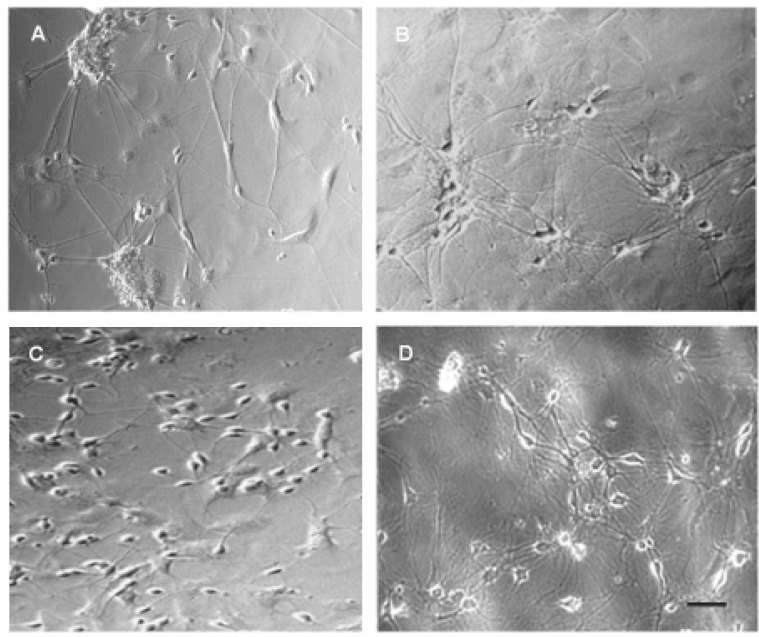
Fig. 1.
Phase contrast microscopy of primary cultured hippocampal (A, B) and cortical (C, D) neurons. (A) Hippocampal neurons of day 4 in vitro. Cultured neurons started neurite sprouting from the early days in vitro. (B) Hippocampal neurons of day 10 in vitro. Neurons are more likely to be aggregated and fully grown neurites shows spider web pattern. (C) Cortical neurons of day 4 in vitro. Neurites are still very short compared to hippocampal neurons of same day in vitro. (D) Cortical neurons of day 13 in vitro. Note that neurites outgrowth of cultured cortical neurons are relatively slow than those of hippocampal neurons. Scale bar is 50 µm.

