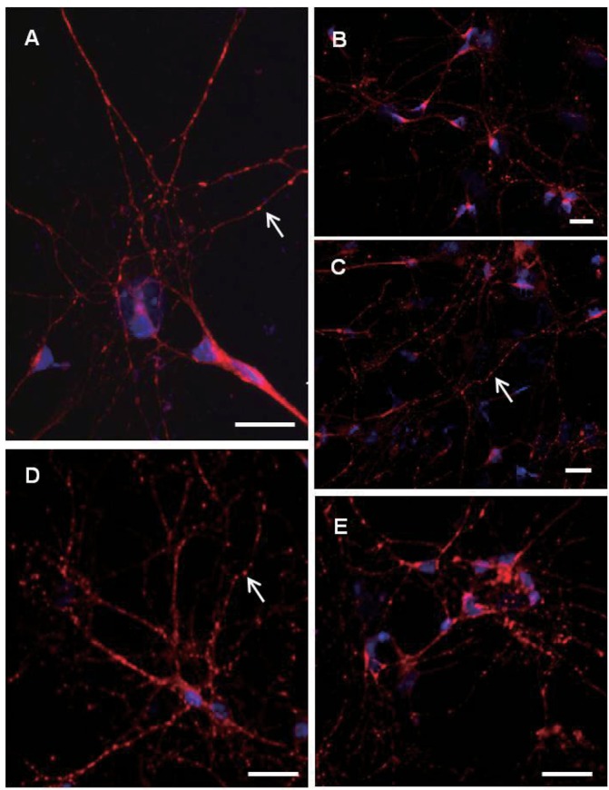
Fig. 3.

Confocal microscopic deteciton of synaptophysin immunopositive cells in pirmary cultured hippocampal neurons at day 2 (A) day 4 (B, C) and day 8 (D, E). Nuclei are counterstained with DAPI (blue color). Note revelation of granular synaptophysin immunofluorescence along the neurites (arrows) from day 2 in vitro with relatively high immunofluorescence levels. Scale bars are 20 µm each.
